Introduction:
Based on multiple wavelength lasers, pinhole, motorized inverted microscope and incubator, confocal laser scanning microscope provides extra high spatial resolution micro-image. This confocal laser scanning microscope can determine direct binding proteins by co-localization or FRET, reconstruct 3D model via high resolution Z-stack micro-images and record physiological response with intensity dynamic change.
It is a powerful tool to study stimulated subcellular target via laser for photo-stimulation and optogenetic application. It is suitable from macroscopic to microscopic observation with different magnification objectives and motor stage for precise mosaic application. Other applications include cell proliferation, cancer therapy, neurite out-growth and long time lapse micro-imaging with stable focusing and incubator for cell viability.
Features:
- Olympus IX-81 motorized inverted fluorescence microscope, motorized focusing with Z-axis drift compensated function.
- Motorized stage for precise positions selection and mosaic micro-imaging.
- Motorized nosepiece and condenser for macro to micro image.
- Stage top incubator for cell viability, 37℃, 5% CO2 and 90% humidity. Injection hole for drug treatment or perfusion system set up.
- Laser wavelength:405nm, 440nm, 488nm, 515nm, 559nm, 635nm.
- With 405nm laser for photo-stimulation via SIM Galvo mirror module.
- Objective:10X, 20X, 40X(Oil), 60X(Oil), 100X(Oil) with DIC.
- Three fluorescence detectors with one DIC detector
Experimental Items:
- High resolution XYZλt and multi-point axis optical section stack.
- Dual laser beam for photo-stimulation.
- Micro-image processing and analysis.
- FRET, FRAP, FLIP, Uncaging, photo-activation, photo-conversion
- Macro to micro with mosaic function.
- Co-localization, physiological dynamic analysis
- Ion influx and out-flux, protein trans-location, cell cycle, proliferation, neurite out-growth, cell interaction, cancer therapy.
Example Data:
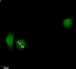
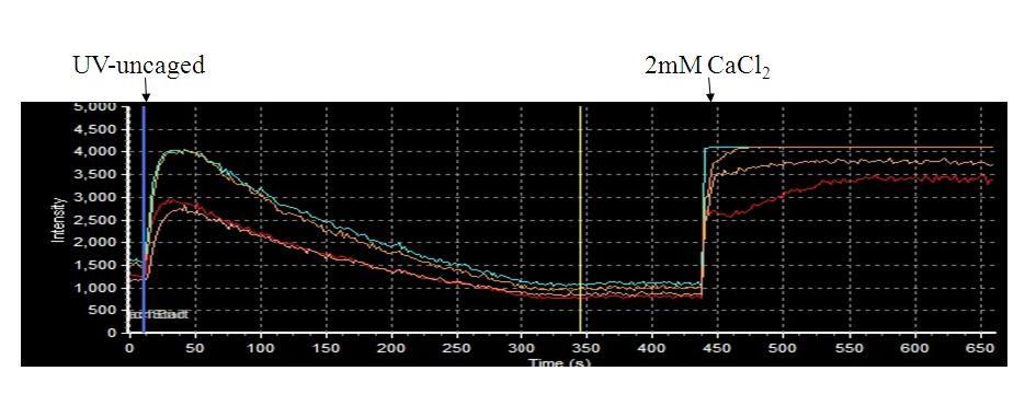
Two beam


Two beam experiment for uncaging application.
One beam

Sample:Huh7 cells
FAQ/Contact:
Name:Chu, Chia-Ying
Tel No.:2371#23
E-mail:cychu@kmu.edu.tw
