
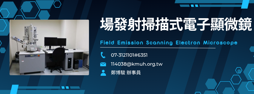
Hitachi SU801 Scanning Electron Microscope (SEM)Hitachi SU801 掃描電子顯微鏡
像以電子束掃描樣品表面,再將二次電子、背向散射電子與 X 光訊號轉成影像及組成資訊:SU801 可觀察細胞、組織、微生物與材料表面,並搭配 EDS 進行元素分析。
01 儀器重點規格 / Key Specifications加速電壓、倍率、解析度、偵測器與樣品尺寸
- 高解析表面影像:提供生物、材料與化學樣品之表面微觀結構觀察。Provides high-resolution surface-microstructure imaging for biological, material, and chemical samples.
- 多偵測器:支援二次電子(SE)與背向散射電子(BSE)偵測。Supports secondary-electron (SE) and backscattered-electron (BSE) detection.
- 元素分析:搭配能量散射 X 光分析(EDS)取得元素組成資訊。Energy-dispersive X-ray spectroscopy (EDS) provides elemental-composition information.
02 核心技術與運作方式 / Core Technology樣品前處理、電子束掃描、SE/BSE 與 EDS
著重樣品表面形貌與微細結構。Secondary-electron imaging emphasizes surface morphology.
呈現不同組成或原子序差異。Backscattered-electron imaging reflects compositional contrast.
能量散射 X 光分析可取得元素資訊。EDS provides elemental-composition data.
電子束聚焦及影像系統可擷取清晰表面結構。Electron-beam focusing and digital imaging capture clear surface structures.
03 研究應用 / Research Applications生物表面、材料微結構、金屬複材與元素分析
觀察生物樣品表面形貌。Examines surface morphology of cells, tissues, and microorganisms.
分析高分子及有機材料之微細結構。Analyzes microstructures of polymers and organic materials.
觀察金屬及複合材料之表面與微結構。Examines surface and microstructure of metals and composites.
以 EDS 取得樣品元素組成。Obtains elemental composition using EDS.
支援材料、化學及跨領域表面分析。Supports materials, chemical, and interdisciplinary surface analysis.
04 分析與數據 / Analysis & Data解析度、倍率、樣品尺寸、多偵測訊號與來源標示
高真空模式解析度標示為 ≤3.0 nm。High-vacuum resolution is specified as ≤3.0 nm.
放大倍率範圍 20X–300,000X。Magnification ranges from 20X to 300,000X.
最大樣品尺寸為直徑 120 mm、高 50 mm。Maximum sample size is 120 mm in diameter and 50 mm in height.
SE、BSE 與 EDS 可分別呈現形貌、組成差異及元素資訊。SE, BSE, and EDS provide morphology, compositional contrast, and elemental information.
原始流程指出完整組織處理約一個月,超過 12 件時依比例延長。The source states full tissue processing takes about one month and longer for more than 12 samples.
05 申請資格與預約流程 / Application & Reservation文件下載、預約、收據、首次諮詢與處理時程
下載電子顯微鏡委託申請表、委託注意事項及研究繳費單。Download the electron-microscopy application form, service guidelines, and payment form.
將申請表與繳費單送至超微細構造研究室並預約。Submit the forms to the Ultrastructural Research Laboratory and schedule service.
於預約時間攜帶繳費單據,經管理者檢視後進行。Bring the payment receipt for verification at the scheduled session.
首次使用或委託檢體製備者,須先溝通安全與樣品需求。First-time users or sample-preparation clients must consult staff in advance.
完整組織處理約一個月;超過 12 件時依比例延長。Allow about one month for full processing, with proportional extension for more than 12 samples.
06 操作規範與注意事項 / Operation Rules繳費、轉帳、收據、首次諮詢、處理時程與退款
醫研部開單後,至啟川大樓 6 樓出納組繳費。After issuance, pay at the Cashier Office on the 6th floor of Chi-Chuan Building.
院內外計畫由財務室轉帳,收款代號 1171。Project transfers use collection code 1171 and are handled by the Finance Office.
預約時須出示繳費單據,經確認後方可實驗。Present the payment receipt for verification before the experiment.
首次使用或送交樣品前處理者須先溝通。First-time users and sample-preparation clients must consult staff.
完整流程約一個月;超過 12 件時依比例延長。Full processing takes about one month and longer for more than 12 samples.
完成收費後無法退費。Fees are non-refundable after payment.
07 使用收費、地點與文件 / Fees, Location & Documents來源所列操作與前處理收費、研究室及相關表單
| 使用者類別/服務 | User Category / Service | 收費標準 |
|---|---|---|
| 院內使用者 | Internal Users | 原始收費欄所列操作費:自行 NT$ 2,400/小時;委託 NT$ 2,800/小時 組織處理委託 NT$ 1,800/小時;超薄切片 NT$ 2,100/塊;離子覆膜自行 NT$ 420/次 |
| 院外使用者 | External Users | 原始收費欄所列操作費:自行 NT$ 4,000/小時;委託 NT$ 4,000/小時 組織處理委託 NT$ 3,000/小時;超薄切片 NT$ 3,500/塊;離子覆膜自行 NT$ 700/次 |
| 育成中心進駐廠商 | Incubation Center Tenant | 原始資料未提供收費 |
醫學研究大樓 2 樓
超微細構造研究室
Ultrastructural Research Laboratory
2nd Floor, Medical Research Building