
應用功能 & 儀器規格
Introduction:
Tissuefaxs® is a unique analytical instrument that combines the advantages of state of the art:
◎ Multi-channel microscopy
◎ Automated high resolution imaging with the scientific accuracy of flow cytometry.
◎ Instead of bleaching away in the fridge, data of entire fluorescence specimens is available on mouse click
to be discussed with your colleagues and students.
◎ The quantitative and objective analysis tools :
Provided by the TissueQuest® software & HistoQuest® software
Module complete the TissueFaxs Performance.
TissueFaxs is successfully used in cancer research, developmental biology, pathology, immunology,
dermatology, urology, drug development and clinical testing.
Function(application):
Microscope-based cell analysis system for cells in tissue sections and smears. TissueFAXS high-throughput acquisition and automated analysis capabilities permit major time savings.
◎ Automated multi-channel image acquisition
◎ Specimen overview and cellular detail in one interface
◎ Individual regions of interest (ROI) definable by graphical tools
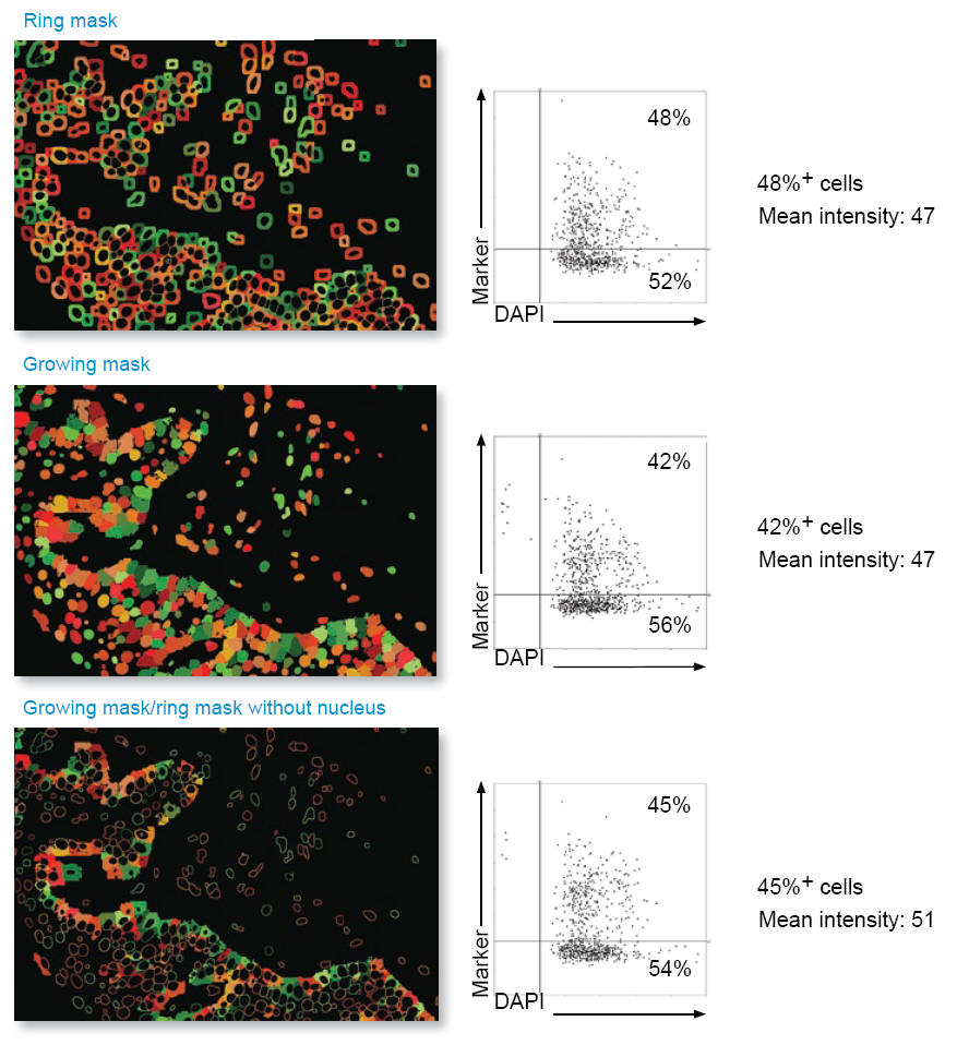
◎ Automated single cell detection by patented algorithms
◎ Objective quantification by control based background subtraction
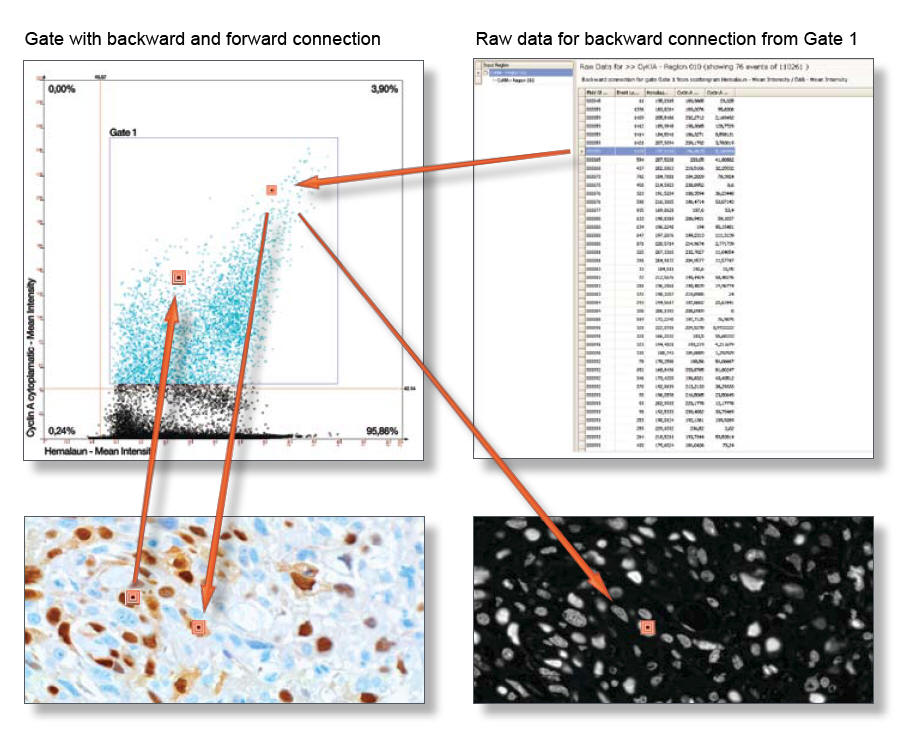
◎ Dotplot operations and gating on subpopulations
◎ Backward and Forward Gating from dotplot to image and image to dotplot
◎ Statistical data in tables and export in Excel as well as ASCII formats
Experimental Items:
The TissueFAXS acquisition and management software combines an intuitive and user friendly interface and a comfortable workflow with extensive control functions:
◎ Objective selection
◎ Light source control
◎ Filter selection
◎ Graphical navigation function for 8 slides
◎ Preview window
◎ ROI management
◎ Well plate layout
◎ Live image window
◎ Stage controls and position info
◎ Autofocus
◎ White balance
◎ Image export
◎ Colour overlay controls
◎ TMA layout


